Laparoscopic Cholecystectomy with ligation of cystic duct by Dr R K Mishra
Add to
Share
683 views
Report
8 months ago
Description
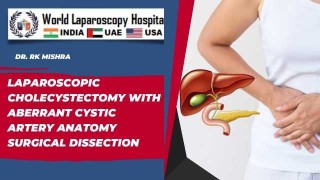
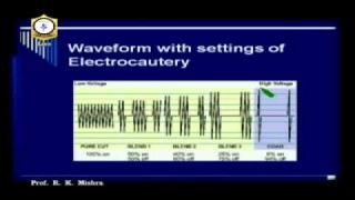
Laparoscopic cholecystectomy has emerged as the gold standard for the surgical treatment of symptomatic gallstone disease. The procedure involves the minimally invasive removal of the gallbladder, offering patients reduced postoperative pain, shorter hospital stays, faster recovery, and minimal scarring compared to open surgery. One of the most crucial steps in this surgery is the safe ligation of the cystic duct, which ensures the prevention of bile leakage and other complications. At World Laparoscopy Hospital, under the expertise of Dr. R. K. Mishra, world-renowned laparoscopic and robotic surgeon, laparoscopic cholecystectomy is performed with the highest standards of precision and safety. Step-by-Step Overview of the Procedure Patient Positioning & Port Placement The patient is placed in the supine position with slight reverse Trendelenburg and left tilt to allow better exposure of the gallbladder. Four trocars are usually inserted: one at the umbilicus for the laparoscope, one in the epigastrium, and two additional working ports in the right hypochondrium. Exposure of Calot’s Triangle Using atraumatic graspers, the gallbladder is retracted to clearly expose the Calot’s triangle. Careful dissection is performed to identify the cystic duct and cystic artery, ensuring the Critical View of Safety (CVS) is obtained before any ligation. Ligation of the Cystic Duct Once the anatomy is clearly defined, the cystic duct is meticulously dissected. Dr. Mishra emphasizes the importance of secure closure to avoid postoperative bile leakage. The duct is ligated either by titanium clips, Hem-o-lok clips, or intracorporeal sutures, depending on the case. Suturing of the cystic duct, performed laparoscopically, is an advanced skill taught at World Laparoscopy Hospital and adds an extra layer of safety in selected patients. Division of the Cystic Duct and Artery After ligation, the cystic duct and cystic artery are divided. Extreme care is taken to avoid injury to the common bile duct, a complication that can have serious consequences if not managed properly. Gallbladder Dissection from Liver Bed The gallbladder is separated from the liver using electrocautery or ultrasonic energy devices. Hemostasis is secured meticulously during this step. Specimen Retrieval The gallbladder is placed in an endo-bag and removed through the umbilical port, ensuring no spillage of bile or stones within the abdominal cavity. Advantages of Dr. Mishra’s Technique Enhanced Safety: Strict adherence to the Critical View of Safety and precise ligation techniques. Teaching Value: Demonstrates advanced skills in cystic duct suturing, a technique particularly useful in cases where clips may be less secure. Minimally Invasive Excellence: Faster recovery, less pain, and minimal scarring. Global Training Impact: Surgeons trained under Dr. Mishra adopt these refined methods, improving surgical outcomes worldwide. Conclusion Laparoscopic cholecystectomy with ligation of the cystic duct is a safe and effective procedure when performed with meticulous technique. Dr. R. K. Mishra, with his vast experience and dedication to advancing minimally invasive surgery, has set a benchmark in performing and teaching this procedure. His contributions not only ensure better patient outcomes but also inspire and train surgeons across the globe to perform laparoscopic surgeries with precision and confidence.
Similar Videos