Laparoscopic Cholecystectomy by Extracorporeal knot
Add to
Share
655 views
Report
8 months ago
Description
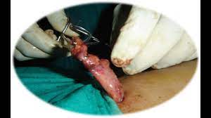
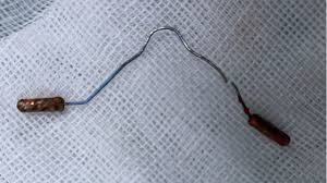
Laparoscopic cholecystectomy is the gold standard procedure for the treatment of gallbladder diseases, including cholelithiasis, cholecystitis, and other biliary disorders. Traditionally, this procedure involves dissecting the gallbladder from the liver bed and ligating the cystic duct and artery using clips or intracorporeal suturing. However, in resource-limited settings or in cases requiring specialized skills, extracorporeal knotting offers an effective and reliable alternative for ligating the cystic duct and artery during laparoscopic cholecystectomy. What is Extracorporeal Knotting? Extracorporeal knotting is a technique in minimally invasive surgery where the suture is tied outside the abdominal cavity and then introduced into the operative field using laparoscopic instruments. Once in place, the knot is slid down to the target site using a knot pusher and secured around the structure, such as the cystic duct or artery. This method avoids the need for complex intracorporeal suturing, making it more accessible for surgeons in the learning phase of laparoscopic surgery. Advantages of Extracorporeal Knot Technique Ease of Learning: It is simpler to learn compared to intracorporeal knotting, reducing the technical barrier for surgeons. Cost-Effective: Eliminates the need for expensive clips or stapling devices. Reliable Ligation: Provides secure closure of the cystic duct and artery, minimizing the risk of postoperative bile leak or hemorrhage. Versatile Application: Can be applied in both elective and emergency cholecystectomy cases. Surgical Technique Patient Positioning: The patient is placed in a supine position with slight reverse Trendelenburg and left tilt to expose the gallbladder. Port Placement: Typically, a 10 mm umbilical port for the camera, a 10 mm epigastric port for instruments, and two 5 mm ports in the right upper quadrant. Dissection: The Calot’s triangle is carefully dissected to identify the cystic duct and cystic artery. Suture Preparation: A non-absorbable or slowly absorbable suture (usually 2-0 or 3-0) is prepared outside the abdomen. A knot is tied extracorporeally. Knot Application: The suture is introduced into the abdominal cavity via a port, looped around the cystic duct or artery, and slid into place using a knot pusher. Additional throws are added if necessary to ensure secure ligation. Gallbladder Removal: After ligation and division of the cystic duct and artery, the gallbladder is dissected from the liver bed using electrocautery and removed through the umbilical port. Clinical Outcomes Studies and surgical experiences have demonstrated that laparoscopic cholecystectomy using extracorporeal knotting is safe, effective, and has comparable outcomes to conventional clip-based techniques. Postoperative complications such as bile leakage, hemorrhage, and infection are minimal when proper technique is followed. Conclusion Laparoscopic cholecystectomy by extracorporeal knot offers a cost-effective, safe, and reliable alternative for gallbladder surgery, especially in settings where clip application may not be feasible. This technique enhances the versatility of minimally invasive surgery and provides surgeons with an additional tool to ensure safe patient outcomes.
Similar Videos