Congenital Müllerian anomalies with two cavitated, non-communicating rudimentary horn
Add to
Share
671 views
Report
8 months ago
Description
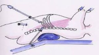
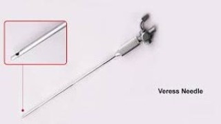
Congenital Müllerian anomalies represent a spectrum of developmental abnormalities arising from defective fusion, resorption, or formation of the paired Müllerian ducts during embryogenesis. These anomalies often present with reproductive, menstrual, or obstetric complications, though some cases remain asymptomatic and are diagnosed incidentally during imaging or surgery. One of the rarest and complex variants is the presence of two cavitated, non-communicating rudimentary horns—an anomaly that poses unique diagnostic and clinical challenges. Embryological Basis During normal embryonic development, the paired Müllerian ducts undergo three essential stages: Organogenesis – formation of both ducts. Fusion – the two ducts fuse to form the uterovaginal canal. Septal resorption – the central septum dissolves, creating a single endometrial cavity. Failure or arrest at different stages results in anomalies. When both ducts fail to develop fully but retain cavitation, two rudimentary horns may form. If these horns are non-communicating, they remain isolated structures, each with its own endometrial cavity but no outflow pathway. Clinical Presentation Patients with cavitated, non-communicating rudimentary horns typically present with: Cyclic pelvic pain (due to outflow obstruction and hematometra). Progressive dysmenorrhea, often worsening with each cycle. Palpable pelvic mass, mimicking ovarian cysts or fibroids. Reproductive difficulties, including infertility, recurrent pregnancy loss, or complications if implantation occurs in a rudimentary horn. In some cases, one horn may be functional while the other is non-functional, further complicating the clinical picture. Diagnostic Challenges Accurate diagnosis is critical but can be difficult. The following modalities are used: Ultrasound (2D/3D) – may demonstrate asymmetric uterine structures but often lacks detail. MRI – the gold standard, allowing precise delineation of uterine anatomy, endometrial cavities, and communication (or lack thereof) between horns. Laparoscopy and hysteroscopy – both diagnostic and therapeutic, confirming the presence of isolated, non-communicating horns. Clinical Implications Obstetric Risks – If pregnancy occurs in a non-communicating rudimentary horn, there is a high risk of ectopic gestation and uterine rupture, often in the second trimester, with catastrophic maternal outcomes. Endometriosis – Retrograde menstruation from a cavitated, obstructed horn can lead to secondary endometriosis. Infertility – Anatomical distortion and recurrent complications may impair fertility. Management Strategies The cornerstone of management is surgical excision of the cavitated, non-communicating rudimentary horn(s). Laparoscopic resection is the preferred approach, offering precise dissection, reduced morbidity, and faster recovery. If both horns are present but one is functional, preserving the functional horn while excising the symptomatic non-communicating one is considered. Fertility counseling is essential, with options such as assisted reproduction discussed when appropriate. Prognosis With timely diagnosis and surgical management, patients generally experience relief from dysmenorrhea, reduced risk of endometriosis, and improved reproductive outcomes. Early recognition is therefore vital, especially in adolescents presenting with cyclic pain soon after menarche. Conclusion Congenital Müllerian anomalies with two cavitated, non-communicating rudimentary horns represent a rare but clinically significant uterine malformation. The condition is associated with severe dysmenorrhea, reproductive complications, and obstetric risks. Advances in imaging, especially MRI, along with minimally invasive surgical techniques, have revolutionized diagnosis and management. A multidisciplinary approach involving gynecologists, radiologists, and reproductive specialists is crucial to optimize outcomes for affected women.
Similar Videos