Fixation of T Tube
Add to
Share
317 views
Report
2 months ago
Description
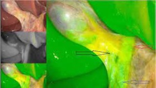
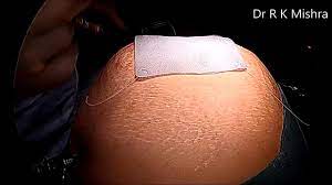
A T-tube is a specialized drainage tube shaped like the letter “T” and is most commonly used after bile duct exploration or choledochotomy. It is placed within the common bile duct to provide decompression, facilitate external drainage of bile, and allow postoperative cholangiography to check for residual stones or strictures. Proper fixation of the T-tube is crucial to prevent dislodgement, leakage of bile into the peritoneal cavity, or accidental removal, which could lead to serious postoperative complications. Indications for T Tube Placement Following open or laparoscopic common bile duct (CBD) exploration After removal of multiple or large CBD stones In cases of choledochoplasty or duct repair When there is significant inflammation or edema of the CBD requiring temporary decompression In high-risk cases where stricture or leak is anticipated Principles of Fixation The fixation of the T-tube involves securing it both intra-abdominally and externally on the skin, ensuring that the tube stays in position while maintaining a proper seal. Intra-abdominal Fixation After placing the horizontal limb of the T-tube inside the CBD, the vertical limb is brought out through a separate stab incision in the abdominal wall, usually in the right upper quadrant. A purse-string suture or interrupted absorbable sutures are placed around the choledochotomy site to snugly fit around the T-tube without compromising bile flow. Care must be taken to avoid kinking of the tube or narrowing of the lumen. External Fixation Once the tube exits the abdominal wall, it should be secured to the skin with non-absorbable sutures (e.g., silk or nylon). A fixation stitch is applied around the tube and tied to the skin to prevent outward migration. The tube is further taped and anchored to the skin to avoid tension or accidental pulling. Ensuring Patency and Security The external end of the tube is connected to a sterile bile collection bag. The tube is flushed gently with normal saline to ensure patency and prevent blockage. Dressing around the exit site must be sterile and changed regularly to prevent infection. Precautions During Fixation Avoid too tight suturing around the CBD to prevent ischemia or stricture. Ensure no bile leakage at the exit site of the tube. Avoid kinking of the T-tube, which can obstruct drainage. Properly label and document the placement to ensure postoperative monitoring. Postoperative Care Monitor bile output and color daily. Perform T-tube cholangiography typically after 10–14 days to check for ductal patency and stone clearance. The T-tube is usually removed after 2–3 weeks, depending on healing and absence of obstruction. Conclusion Fixation of a T-tube is a critical surgical step that ensures safe decompression of the biliary system after common bile duct exploration. Secure intra-abdominal placement, reinforced external fixation, and diligent postoperative monitoring minimize complications such as dislodgement, bile leak, and infection, ultimately contributing to a smooth recovery and favorable patient outcomes.
Similar Videos


