Laparoscopic Appendectomy: Anatomy and Step-by-Step Appendix Removal Procedure
Add to
Share
10 views
Report
14 hours ago
Description
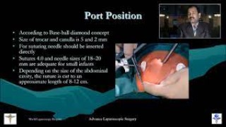
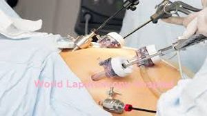
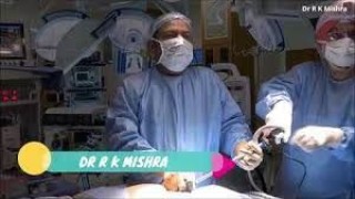
Laparoscopic Appendectomy is one of the most commonly performed minimally invasive surgeries worldwide. At World Laparoscopy Hospital (WLH), this procedure is taught and performed with the highest precision using advanced laparoscopic techniques, ensuring minimal pain, faster recovery, and superior cosmetic results for patients. Under the guidance of Dr. R. K. Mishra, a pioneer in laparoscopic surgery, surgeons at WLH master the complete anatomical understanding and surgical skills needed to perform safe and effective appendectomies. Anatomy of the Appendix The vermiform appendix is a narrow, worm-shaped tube connected to the cecum—the beginning of the large intestine. It is typically located in the right lower quadrant of the abdomen, known as the right iliac fossa. Length: 5–10 cm Position Variations: Retrocecal (most common), pelvic, subcecal, preileal, and postileal Blood Supply: Appendicular artery (a branch of the ileocolic artery) Function: Though once considered vestigial, it is now known to play a role in immune function, especially in young individuals. Understanding the variable anatomy of the appendix is critical for laparoscopic surgeons to identify and remove it safely without injury to nearby structures such as the cecum or ileum. Indications for Laparoscopic Appendectomy Acute appendicitis Recurrent or chronic appendicitis Appendiceal abscess (after initial conservative management) Diagnostic uncertainty (when appendicitis is suspected but not confirmed) At WLH, surgeons also learn how to handle complicated cases such as perforated appendicitis or appendicitis with peritonitis using advanced laparoscopic techniques. Step-by-Step Laparoscopic Appendix Removal Procedure 1. Patient Preparation The patient is placed under general anesthesia and positioned supine with a slight Trendelenburg tilt and left tilt to move the small bowel away from the operative field. After aseptic preparation, a Foley catheter and nasogastric tube are often inserted. 2. Port Placement Typically, three ports are used: 10 mm umbilical port – for the camera 5 mm suprapubic port – for the surgeon’s left hand 5 mm left lower quadrant port – for the working instrument in the right hand This triangular configuration provides optimal visualization and instrument maneuverability. 3. Diagnostic Laparoscopy The surgeon first inspects the entire abdomen to confirm the diagnosis and rule out other causes of pain such as pelvic inflammatory disease or Meckel’s diverticulum. 4. Identification of the Appendix The cecum is traced to locate the base of the appendix. The surgeon carefully exposes the appendix by retracting it and dissecting the mesoappendix, which contains the appendicular artery. 5. Skeletonization and Division The mesoappendix is divided using bipolar cautery, harmonic scalpel, or advanced energy devices, ensuring proper hemostasis. The appendicular artery is ligated securely. 6. Ligation and Resection The base of the appendix is tied with two or three endoloops (or stapled using an endoscopic stapler). The appendix is then transected and placed in a retrieval bag to prevent contamination during extraction. 7. Inspection and Irrigation The appendiceal stump and surrounding area are inspected for bleeding or leakage. The peritoneal cavity is irrigated with warm saline if required. 8. Specimen Removal and Closure The specimen is removed through the umbilical port, and all ports are closed carefully to prevent herniation. Cosmetic results are excellent, with barely visible scars. Advantages of Laparoscopic Appendectomy at WLH Minimal postoperative pain Early return to normal activities Shorter hospital stay Reduced wound infection rates Excellent cosmetic results Superior 3D high-definition visualization during surgery World Laparoscopy Hospital’s facilities are equipped with state-of-the-art laparoscopic systems, 3D vision, and artificial intelligence-assisted simulators, providing real-time training to surgeons from over 108 countries. Training Excellence at World Laparoscopy Hospital At WLH, laparoscopic appendectomy is not only performed for patient care but also serves as a teaching model for surgical education. Trainee surgeons practice the full step-by-step procedure on animal models, virtual reality simulators, and live surgeries under expert supervision. Through Fellowship and Diploma courses in Minimal Access Surgery, participants gain hands-on experience and confidence to perform appendectomy and other advanced laparoscopic surgeries independently. Conclusion Laparoscopic Appendectomy at World Laparoscopy Hospital combines advanced technology, anatomical mastery, and surgical expertise. The minimally invasive approach ensures patient safety, faster recovery, and outstanding outcomes while maintaining the highest standards of surgical education for future laparoscopic surgeons.
Similar Videos