Splenic Ischemia: A Rare Complication of Laparoscopic Nissen Fundoplication
Add to
Share
584 views
Report
7 months ago
Description
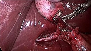
Laparoscopic Nissen fundoplication is a gold-standard surgical procedure for treating gastroesophageal reflux disease (GERD). This minimally invasive operation aims to restore the function of the lower esophageal sphincter by wrapping the gastric fundus around the esophagus. One of the critical intraoperative steps often involves dividing the short gastric vessels to achieve adequate mobilization of the gastric fundus. However, in rare cases, this maneuver can lead to an uncommon but significant complication — splenic ischemia. At World Laparoscopy Hospital (WLH), where advanced minimally invasive surgery and surgical education meet global standards, such cases are handled with utmost precision and serve as valuable learning experiences for surgeons worldwide. Under the expert guidance of Dr. R. K. Mishra, the hospital has extensively studied and documented complex scenarios like splenic ischemia following short gastric vessel division, turning each case into an opportunity for surgical excellence and teaching. Understanding the Complication Splenic ischemia occurs when blood flow to part of the spleen becomes compromised, often due to inadvertent damage to the splenic arterial branches or excessive traction on the short gastric vessels during dissection. Although rare, the condition can result in localized infarction of splenic tissue, which may present as postoperative pain in the left upper quadrant, fever, or elevated inflammatory markers. Why It Happens During a standard Nissen fundoplication, surgeons may choose to divide the short gastric vessels to ensure a tension-free fundic wrap. However, this division must be performed with careful identification of vascular anatomy to avoid compromising the splenic hilum or the posterior gastric artery. In some patients, anatomical variations make the spleen more susceptible to ischemic injury when these vessels are divided too close to the splenic capsule or if energy devices are misapplied. Diagnosis and Management At WLH, the diagnosis of splenic ischemia is achieved through high-resolution laparoscopic visualization during surgery and postoperative imaging such as contrast-enhanced CT scans. Most cases of minor ischemia resolve conservatively with close monitoring, pain management, and antibiotics if needed. However, significant ischemia or abscess formation may require percutaneous drainage or, rarely, splenectomy. The hospital’s structured postoperative surveillance protocol ensures that patients are monitored for early signs of complications, allowing timely intervention. This reflects WLH’s philosophy — “Safety through skill, precision, and vigilance.” Educational Value for Surgeons One of the distinguishing aspects of World Laparoscopy Hospital is its emphasis on training surgeons in recognizing and preventing such complications. During hands-on sessions in the Center of Excellence for Minimal Access Surgery, trainees learn: The vascular anatomy of the gastric fundus and spleen in detail. Safe dissection techniques for dividing short gastric vessels. Strategies for maintaining hemostasis and avoiding traction injuries. Real-case scenario discussions on splenic ischemia management. These insights transform a potential complication into an invaluable educational moment, reinforcing WLH’s commitment to excellence in laparoscopic teaching and patient safety. Conclusion While splenic ischemia after short gastric vessel division during laparoscopic Nissen fundoplication is an uncommon event, it underscores the importance of meticulous surgical technique, anatomical knowledge, and intraoperative vigilance. The experience at World Laparoscopy Hospital exemplifies how advanced technology, expert mentorship, and a culture of continuous learning can prevent, identify, and effectively manage such rare complications.
Similar Videos