Understanding eTEP Anatomy: Landmarks for Safe Hernia Repair
Add to
Share
1,517 views
Report
7 months ago
Description
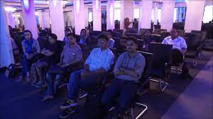
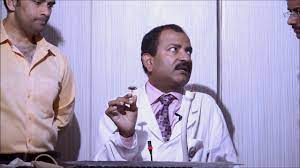
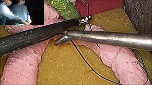
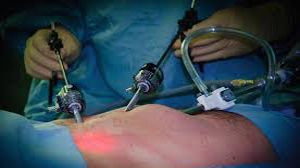
At World Laparoscopy Hospital, surgeons are redefining precision in hernia surgery through the Extended Totally Extraperitoneal (eTEP) approach—an evolution in minimally invasive inguinal hernia repair. This advanced technique emphasizes a deep understanding of abdominal wall anatomy, meticulous dissection, and the identification of key anatomical landmarks to ensure safe and effective outcomes. Understanding the eTEP Concept The eTEP technique expands the traditional totally extraperitoneal (TEP) approach by creating a wider working space that extends beyond the midline, allowing for bilateral access and flawless visualization of myopectineal orifices. Unlike the conventional TEP, eTEP provides ergonomic access for the surgeon, less instrument crowding, and better anatomical orientation—all critical for teaching and performing high-quality repairs at World Laparoscopy Hospital. Key Anatomical Landmarks in eTEP Inguinal Hernia Repair Linea Alba and Rectus Abdominis Muscle The dissection begins by identifying the linea alba and entering the retrorectus space. Preserving the posterior rectus sheath is crucial, as it forms the initial working cavity for developing the extraperitoneal plane. Arcuate Line (Linea Semicircularis) The arcuate line marks the transition where the posterior rectus sheath ends. Below this point, the peritoneum is directly related to the transversalis fascia—an important consideration while expanding the preperitoneal space to avoid peritoneal breach. Epigastric Vessels The inferior epigastric vessels serve as vital guides separating the direct and indirect hernia spaces. Preserving them ensures minimal bleeding and helps in identifying the lateral compartment during mesh placement. Cooper’s Ligament (Pectineal Ligament) A solid fibrous structure along the superior pubic ramus, Cooper’s ligament is a key anchoring point for mesh fixation. It defines the medial boundary of the myopectineal orifice and helps orient the surgeon to the Hesselbach’s triangle. Hesselbach’s Triangle Bounded by the inferior epigastric vessels laterally, the rectus abdominis medially, and the inguinal ligament inferiorly, this is the zone for direct hernia defects. Accurate identification ensures proper dissection and mesh overlap to prevent recurrence. Spermatic Cord Structures The vas deferens and testicular vessels pass through the deep ring laterally and should be carefully preserved. Proper identification and skeletonization of these structures are crucial to prevent chronic pain or testicular ischemia. Myopectineal Orifice of Fruchaud This is the central region of hernia vulnerability encompassing all possible hernia sites—direct, indirect, and femoral. The eTEP approach provides a panoramic view of this orifice, allowing for comprehensive reinforcement with a single mesh. Peritoneal Reflection and Dissection Planes Gentle dissection along the peritoneal reflection is essential to maintain a large, smooth working field. Avoiding peritoneal tears ensures clear vision, stable pneumopreperitoneum, and easier bilateral exploration. Educational Excellence at World Laparoscopy Hospital At World Laparoscopy Hospital (WLH), surgeons are trained under the direct guidance of Dr. R. K. Mishra, an internationally renowned laparoscopic expert. The eTEP training module focuses not only on technical mastery but also on three-dimensional anatomical understanding. High-definition 3D models, simulation labs, and live operative sessions help trainees gain confidence in recognizing and preserving these vital landmarks during real-time surgery. Conclusion Mastering eTEP inguinal hernia anatomy is the cornerstone of achieving excellence in minimally invasive repair. By combining anatomical precision, innovative technique, and world-class mentorship, World Laparoscopy Hospital continues to shape the next generation of surgeons who perform with accuracy, safety, and superior patient outcomes.
Similar Videos