Managing a Wide Cystic Duct: Laparoscopic Cholecystectomy Technique
Add to
Share
915 views
Report
7 months ago
Description
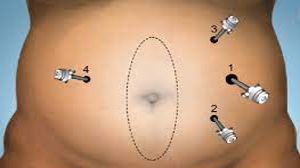
Laparoscopic cholecystectomy has become the gold standard for the management of symptomatic gallstones and chronic cholecystitis. However, anatomical variations such as an unusually wide cystic duct can make the procedure technically challenging, even for experienced surgeons. At World Laparoscopy Hospital (WLH), surgeons are trained to manage such complex scenarios with precision, safety, and minimal invasiveness. Understanding the Challenge The cystic duct usually measures around 2–4 mm in diameter, but in certain cases—especially in chronic inflammation or biliary obstruction—it may become significantly dilated, sometimes exceeding 10 mm. A wide cystic duct poses difficulties in clip application, risk of bile leak, or inadvertent common bile duct injury if not properly identified and managed. Preoperative Assessment At WLH, every laparoscopic cholecystectomy begins with a meticulous preoperative workup. Ultrasonography or MRCP (Magnetic Resonance Cholangiopancreatography) helps evaluate the size of the cystic duct and detect any anatomical variations or associated stones. Patients are counseled about the possibility of advanced intraoperative techniques, including intracorporeal suturing or endoloop ligation, if clipping is not feasible. Step-by-Step Surgical Technique 1. Port Placement and Access A standard four-port technique is employed: A 10 mm umbilical port for the camera. A 10 mm epigastric working port. Two 5 mm ports in the right subcostal area for traction and dissection. The abdomen is insufflated with CO₂ to create an optimal working space. 2. Exposure and Retraction The fundus of the gallbladder is retracted cranially, and the infundibulum is pulled laterally to open Calot’s triangle. This step provides a clear view of the cystic duct and artery. 3. Dissection of Calot’s Triangle Gentle dissection using Maryland forceps and energy sources (such as harmonic scalpel or bipolar) is carried out. The cystic duct is identified, and the Critical View of Safety (CVS) is achieved — a mandatory principle followed at WLH to prevent bile duct injuries. 4. Identification of the Wide Cystic Duct When the cystic duct appears unusually wide, its size and course are carefully assessed. Traction is adjusted to avoid false dissection. Intraoperative cholangiography may be performed to confirm anatomy if there is any uncertainty. 5. Safe Closure of the Wide Cystic Duct Depending on the duct diameter and wall thickness, surgeons at WLH use different advanced methods: Endoloop Ligation (PDS or Vicryl loop) for ducts too wide for standard clips. Intracorporeal Suturing with absorbable 2-0 Vicryl for very large or friable ducts. Large Titanium or Hem-o-lok Clips when the duct can still be securely occluded. The closure technique is selected to ensure complete ductal sealing and prevent postoperative bile leakage. 6. Division of the Cystic Duct and Artery After securing the duct, it is carefully divided with laparoscopic scissors. The cystic artery is clipped and divided in a standard manner, maintaining constant awareness of the nearby common bile duct. 7. Gallbladder Separation from Liver Bed Using an energy device, the gallbladder is dissected from the hepatic bed with minimal bleeding. Continuous suction and irrigation maintain a clean operative field. 8. Specimen Retrieval and Final Inspection The gallbladder is extracted through the epigastric port in an endo-bag. The operative site is inspected for any bile leak or bleeding. A drain may be placed selectively. Postoperative Care and Recovery Patients typically recover quickly with minimal pain and are discharged within 24–48 hours. At WLH, postoperative care includes early ambulation, oral intake after 6 hours, and ultrasound follow-up if needed. Learning Excellence at World Laparoscopy Hospital Surgeons trained at WLH are exposed to a wide variety of real and simulated biliary cases, including complex cystic duct variations. Through hands-on training, high-definition video lectures, and mentorship under Dr. R. K. Mishra, trainees learn not only the technical steps but also the judgment required for safe decision-making in challenging anatomical situations. Conclusion Managing an unusually wide cystic duct during laparoscopic cholecystectomy demands a refined surgical technique, deep anatomical understanding, and careful intraoperative strategy. At World Laparoscopy Hospital, the combination of advanced technology and expert guidance ensures that even the most complex gallbladder surgeries are performed with confidence, precision, and patient safety at the forefront.
Similar Videos