Safe Laparoscopic Cholecystectomy in Acute Cholecystitis
Add to
Share
597 views
Report
5 months ago
Description
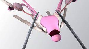
Acute cholecystitis is a common surgical emergency, most often resulting from obstruction of the cystic duct by gallstones, leading to inflammation, edema, and infection of the gallbladder. Laparoscopic cholecystectomy has become the gold standard for the management of acute cholecystitis due to its advantages of reduced postoperative pain, shorter hospital stay, faster recovery, and better cosmetic outcomes. However, surgery in the acute setting demands meticulous technique, particularly during cystic duct ligation, to avoid bile duct injury. The inflamed gallbladder in acute cholecystitis is frequently distended, friable, and surrounded by dense adhesions. Initial steps include careful adhesiolysis and decompression of the gallbladder when required, to improve exposure. Retraction of the gallbladder fundus towards the right shoulder and lateral traction on the infundibulum helps to open Calot’s triangle. Dissection should always be performed close to the gallbladder wall, using blunt and sharp techniques, to minimize the risk of injury to adjacent structures. Cystic duct ligation is a critical step in laparoscopic cholecystectomy. The cystic duct should be clearly identified, skeletonized, and differentiated from the common bile duct. In acute cholecystitis, the cystic duct may be short, edematous, or wide, making identification challenging. Once safely exposed, the duct is usually secured with titanium clips, locking clips, or intracorporeal sutures depending on its size and condition. Adequate spacing between clips and careful division of the duct are essential to prevent postoperative bile leak. The concept of the Critical View of Safety (CVS) is fundamental in preventing bile duct injuries, especially in difficult cases of acute inflammation. CVS requires three essential criteria to be met before division of any tubular structure: first, the hepatocystic triangle must be completely cleared of fat and fibrous tissue; second, the lower one-third of the gallbladder must be dissected off the liver bed; and third, only two structures—the cystic duct and cystic artery—should be seen entering the gallbladder. Achieving CVS confirms correct anatomical identification and significantly reduces the risk of misidentification injuries. In conclusion, laparoscopic cholecystectomy in acute cholecystitis is safe and effective when performed with adherence to sound surgical principles. Careful dissection, secure cystic duct ligation, and strict achievement of the Critical View of Safety are the cornerstones of a successful procedure. When anatomy is unclear, surgeons should not hesitate to adopt alternative strategies such as subtotal cholecystectomy or conversion to open surgery, as patient safety remains the highest priority.
Similar Videos