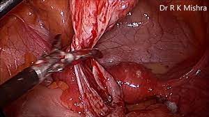
Dr. Mishra's Lecture on Female Pelvic Anatomy at CAMLS, Florida, USA
Add to
Share
2,663 views
Report
3 years ago
Description
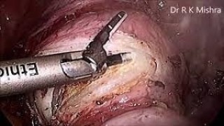
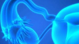
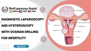
Dr. R. K. Mishra's Lecture on Female Pelvic Anatomy at CAMLS, University of South Florida, USA. Fellowship of Minimal Acess Surgery Training course conducted by World Laparoscopy Training Institute, Florida, United States of America. The pelvis is the lower part of the torso. It’s located between the abdomen and the legs. This area provides support for the intestines and also contains the bladder and reproductive organs. There are some structural differences between the female and the male pelvis. Most of these differences involve providing enough space for a baby to develop and pass through the birth canal of the female pelvis. As a result, the female pelvis is generally broader and wider than the male pelvis. Female pelvis organs Uterus The uterus is a thick-walled, hollow organ where a baby develops during pregnancy. During the reproductive years, the lining of the uterus sheds every month during menstruation if you don’t become pregnant. Ovaries There are two ovaries located on either side of the uterus. The ovaries produce eggs and also release hormones, such estrogen and progesterone. Fallopian tubes The fallopian tubes connect each ovary to the uterus. Specialized cells in the fallopian tubes use hair-like structures called cilia to help direct eggs from the ovaries toward the uterus. Cervix The cervix connects the uterus to the vagina. It’s able to widen, allowing sperm to pass into the uterus. In addition, thick mucus produced in the cervix can help to prevent bacteria from reaching the uterus. Vagina The vagina connects the cervix to the exterior female genitalia. It’s also called the birth canal, as the baby passes through the vagina during delivery. Rectum The rectum is the lowest part of the large intestine. Feces collects here until exiting through the anus. Bladder The bladder is the organ that collects and stores urine until it’s released. Urine reaches the bladder through tubes called ureters that connect to the kidneys. Urethra The urethra is the tube that urine travels through to exit the body from the bladder. The female urethra is much shorter than the male urethra. Female pelvis ligaments Broad ligament The broad ligament supports the uterus, fallopian tubes, and ovaries. It extends to both sides of the pelvic wall. The broad ligament can be further divided into three components that are linked to different parts of the female reproductive organs: mesometrium, which supports the uterus mesovarium, which supports the ovaries mesosalpinx, which supports the fallopian tubes Uterine ligaments Uterine ligaments provide additional support for the uterus. Some of the main uterine ligaments include: the round ligament cardinal ligaments pubocervical ligaments uterosacral ligaments Ovarian ligaments The ovarian ligaments support the ovaries. There are two main ovarian ligaments: the ovarian ligament the suspensory ligament of the ovary https://www.laparoscopyhospital.com/ For more information please contact: World Laparoscopy Hospital Cyber City, Gurugram, NCR DELHI INDIA 122002 Phone & WhatsApp: +919811416838, + 91 9999677788
Similar Videos