Laparoscopic IPOM for Ventral Hernia: Step-by-Step Repair Guide
Add to
Share
2,260 views
Report
7 months ago
Description
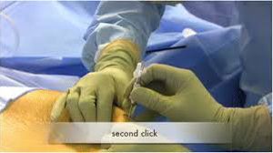
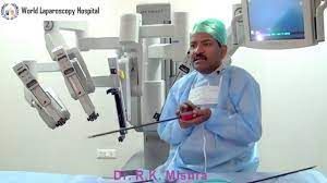
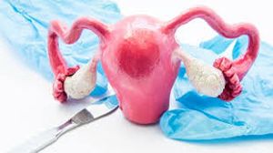
The Intraperitoneal Onlay Mesh (IPOM) technique has revolutionized the management of ventral hernias, offering a minimally invasive, tension-free, and cosmetically superior solution compared to traditional open repair. At World Laparoscopy Hospital (WLH), under the expert guidance of Dr. R. K. Mishra, the IPOM technique is taught and performed with the highest precision, emphasizing safety, anatomical understanding, and advanced laparoscopic skill. Overview of IPOM Technique Laparoscopic IPOM repair involves placing a specially designed composite mesh on the inner surface of the abdominal wall to cover the hernia defect from within the peritoneal cavity. The mesh provides strong reinforcement, preventing recurrence and minimizing postoperative pain and infection. Step-by-Step Surgical Procedure 1. Patient Preparation and Positioning The patient is placed in a supine position under general anesthesia. The abdomen is prepared and draped in a sterile fashion. A Foley catheter may be inserted for bladder decompression. The operating surgeon typically stands on the patient’s left side with the monitor positioned opposite for optimal visualization. 2. Port Placement and Pneumoperitoneum A Veress needle or open (Hasson) technique is used to establish pneumoperitoneum. Three to four trocars are strategically placed depending on the location of the hernia: 10 mm umbilical port for the camera. 5 mm or 10 mm working ports on either side of the abdomen. The key is to maintain triangulation for efficient dissection and mesh deployment. 3. Diagnostic Laparoscopy and Adhesiolysis A thorough inspection of the abdominal cavity is performed. Adhesions around the hernia sac are meticulously released using energy devices (harmonic scalpel or bipolar). This step requires precision to avoid bowel injury, especially in recurrent or post-surgical hernias. 4. Hernia Sac Reduction The hernia contents—often omentum or bowel loops—are carefully reduced back into the peritoneal cavity. The defect margins are cleaned, ensuring a clear field for mesh placement. 5. Defect Closure (Optional but Recommended) Many surgeons at WLH advocate primary closure of the defect using non-absorbable sutures. This restores the abdominal wall contour and prevents mesh bulging (IPOM-plus technique). 6. Mesh Selection and Preparation A composite dual-layer mesh is used—one side designed for tissue ingrowth and the other coated to prevent bowel adhesion. The mesh is tailored to extend at least 3–5 cm beyond the defect margins in all directions. 7. Mesh Introduction and Fixation The mesh is rolled and inserted through the 10 mm trocar. Once unfolded and positioned, it is secured using transfascial sutures and absorbable tackers in a “double crown” configuration: Inner circle for initial anchoring. Outer circle for reinforcement and tension-free fixation. This ensures durable repair and minimizes mesh migration. 8. Hemostasis and Desufflation After mesh placement, the field is inspected for hemostasis. Pneumoperitoneum is released gradually, and the trocars are removed under direct vision. Skin incisions are closed with absorbable sutures for an aesthetic finish. Postoperative Care Patients typically experience minimal pain and recover rapidly. Early ambulation is encouraged within a few hours after surgery. Oral intake resumes on the same day, and discharge often occurs within 24–48 hours. Return to normal activities is possible within a week, emphasizing the minimally invasive nature of IPOM repair. Advantages of IPOM Repair at WLH Minimal tissue trauma and faster recovery Lower recurrence rates due to optimal mesh overlap and fixation Excellent cosmetic results with small incisions World-class training environment ensuring hands-on skill development under expert supervision Use of state-of-the-art 3D laparoscopic systems and energy devices Training and Excellence at World Laparoscopy Hospital At World Laparoscopy Hospital, Gurgaon, Dubai, and USA, surgeons from across the globe master the IPOM technique through intensive hands-on training, live surgeries, and simulation labs. The curriculum, led by Dr. R. K. Mishra, emphasizes not only surgical skill but also ergonomics, complication management, and evidence-based best practices. Conclusion The Laparoscopic IPOM Ventral Hernia Repair represents the perfect blend of surgical innovation and patient safety. At World Laparoscopy Hospital, this procedure is not just a surgery—it’s a demonstration of minimally invasive excellence, refined technique, and the commitment to advancing laparoscopic education worldwide.
Similar Videos