Step-by-Step Laparoscopic Repair of Pfannenstiel Incisional Hernia After Cesarean Section
Add to
Share
730 views
Report
3 months ago
Description
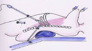
Pfannenstiel incision, commonly used for cesarean section, is preferred for its cosmetic advantage and reduced postoperative discomfort. However, in rare cases, patients may develop an incisional hernia at the site of this transverse suprapubic incision. A Pfannenstiel incisional hernia after cesarean section can lead to pain, swelling, bowel-related symptoms, and reduced quality of life. At World Laparoscopy Hospital, the laparoscopic repair of such hernias is performed with precision, following a standardized, evidence-based step-by-step protocol that ensures safety, minimal trauma, and excellent recovery outcomes. Preoperative Assessment and Preparation The management begins with a detailed clinical evaluation. Patients typically present with a swelling in the lower abdomen, which becomes more prominent on coughing or straining. Ultrasonography or contrast-enhanced CT scan helps confirm the diagnosis, determine the size of the defect, and evaluate the contents of the hernia sac. Preoperative optimization includes control of comorbidities such as diabetes, obesity, or anemia. Prophylactic antibiotics are administered before incision, and the patient is placed under general anesthesia. Bladder catheterization is performed to avoid injury during trocar placement, especially considering the proximity of the defect to the suprapubic region. Patient Positioning and Port Placement The patient is placed in the supine position with slight Trendelenburg tilt to allow bowel loops to fall away from the pelvis. After establishing pneumoperitoneum using a Veress needle or open (Hasson) technique, a 10 mm camera port is inserted, usually at the umbilicus or slightly above it. Two additional 5 mm working ports are placed laterally under direct vision, ensuring they are positioned away from the hernia defect to provide optimal triangulation and ergonomics. Proper port placement is crucial to achieve effective dissection and mesh placement. Diagnostic Laparoscopy and Adhesiolysis Once the laparoscope is introduced, a thorough diagnostic laparoscopy is performed. The hernia defect at the Pfannenstiel incision site is identified. Adhesions between the omentum, bowel, and anterior abdominal wall are common in post-cesarean patients. Meticulous adhesiolysis is performed using atraumatic graspers and energy devices. Care is taken to prevent bowel injury, as the lower abdominal location may involve adhesions close to the urinary bladder. Complete reduction of hernia contents into the abdominal cavity is achieved before proceeding further. Identification and Measurement of the Defect After reduction of contents, the fascial defect is clearly visualized. The size of the hernia defect is measured using a sterile measuring scale or by external palpation and internal visualization. Accurate measurement is essential to select an appropriately sized mesh that provides at least 3–5 cm overlap beyond the margins of the defect in all directions. Closure of the Defect (IPOM Plus Technique) At World Laparoscopy Hospital, surgeons often prefer the IPOM Plus (Intraperitoneal Onlay Mesh with defect closure) technique. The fascial defect is closed intracorporeally using non-absorbable or delayed absorbable sutures. Closure of the defect restores the anatomy, reduces seroma formation, and enhances abdominal wall strength. Mesh Placement and Fixation A composite mesh designed for intraperitoneal placement is introduced into the abdominal cavity through the 10 mm port. The mesh is carefully unrolled and positioned over the defect, ensuring adequate overlap on all sides. Mesh fixation is achieved using a combination of transfascial sutures and absorbable or non-absorbable tackers. Particular attention is given to avoid injury to the inferior epigastric vessels and the urinary bladder. Proper fixation prevents recurrence and ensures long-term durability of the repair. Final Inspection and Closure After securing the mesh, the operative field is inspected for hemostasis. Pneumoperitoneum is released gradually while observing the mesh position to ensure it remains properly placed. Ports are removed under direct vision, and the fascia at the 10 mm port site is closed to prevent port-site hernia. Skin incisions are closed with absorbable sutures for optimal cosmetic results. Postoperative Care and Recovery Patients undergoing laparoscopic repair typically experience minimal postoperative pain compared to open surgery. Early ambulation is encouraged within hours of surgery. Most patients are discharged within 24–48 hours, depending on their clinical condition. Follow-up includes wound assessment, monitoring for seroma, and evaluation of return to normal activities. Patients are advised to avoid heavy lifting for several weeks to allow adequate healing. Advantages of Laparoscopic Repair The laparoscopic approach offers several benefits: Smaller incisions and improved cosmetic outcomes Reduced postoperative pain Lower risk of wound infection Faster recovery and early return to daily activities Better visualization of the defect and surrounding structures Conclusion Step-by-step laparoscopic repair of Pfannenstiel incisional hernia after cesarean section represents a safe, effective, and minimally invasive solution. With structured training, advanced technology, and adherence to standardized protocols, World Laparoscopy Hospital continues to deliver excellence in hernia surgery. The combination of precise surgical technique, careful patient selection, and comprehensive postoperative care ensures durable results and high patient satisfaction.
Similar Videos