3-Port Laparoscopic Cholecystectomy with ICG Fluorescence by Dr. R.K. Mishra
Add to
Share
5 views
Report
6 hours ago
Description
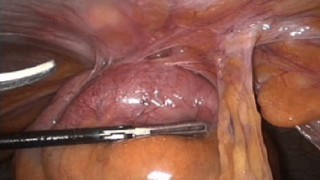
Discover the principles of three-port laparoscopic cholecystectomy with indocyanine green (ICG) fluorescence imaging in this educational surgical video from World Laparoscopy Hospital (WLH). This demonstration explores the combination of a minimally invasive gallbladder removal technique with fluorescence-guided visualization of the biliary anatomy, offering valuable learning for general surgeons, laparoscopic surgery trainees, and medical professionals interested in advanced minimal access surgery. Laparoscopic cholecystectomy is a widely performed procedure for treating symptomatic gallbladder disease, including gallstones. The three-port approach uses a camera and two additional working ports, with instrument positioning and operative strategy adapted to the patient's anatomy and the surgeon's preferred technique. When suitable equipment is available, ICG fluorescence cholangiography can provide real-time visualization of extrahepatic biliary structures during the operation. Understanding Three-Port Laparoscopic Cholecystectomy Traditional laparoscopic cholecystectomy commonly uses four ports. A three-port approach reduces the number of abdominal access sites and requires coordinated camera control, effective traction, and careful instrument manipulation. The operative strategy must maintain adequate exposure and allow the surgeon to identify the relevant anatomy before dividing any structures. The number of ports alone does not determine surgical safety. Appropriate patient selection, adequate visualization, sound anatomical judgment, and the ability to change the operative approach when necessary remain essential. What Is ICG Fluorescence Imaging? Indocyanine green is a fluorescent dye that emits near-infrared light when illuminated by a compatible imaging system. Following intravenous administration, ICG is taken up by the liver and excreted into bile. A fluorescence-enabled laparoscopic camera can then help visualize biliary structures in real time. During cholecystectomy, fluorescence imaging may help identify the cystic duct and other extrahepatic biliary structures before and during dissection. Evidence and professional consensus suggest that ICG fluorescence can improve anatomical visualization, although its effectiveness varies with inflammation, tissue thickness, and the individual patient's anatomy. Step-by-Step Overview of the Surgical Technique 1. Preoperative preparation The surgical team evaluates the indication for cholecystectomy, relevant medical history, imaging findings, and anesthetic requirements. The planned use of ICG is reviewed according to the patient's circumstances, local protocols, and any relevant contraindications or previous adverse reactions. 2. Establishing three-port access Under general anesthesia, the surgeon establishes laparoscopic access and positions the camera and working ports to provide suitable visualization and instrument movement. The placement of ports depends on patient anatomy, the location of the gallbladder, and the intended operative technique. 3. Initial inspection and exposure The surgeon examines the gallbladder and surrounding anatomy, assessing inflammation, adhesions, and technical difficulty. Appropriate traction and countertraction expose the hepatocystic triangle and help establish a clear operative field. 4. Fluorescence-guided biliary visualization With a compatible near-infrared imaging system, the surgeon assesses the fluorescent biliary anatomy. Imaging may be reviewed before dissection, during dissection of the hepatocystic triangle, and whenever additional anatomical information is needed. The fluorescence view complements conventional laparoscopic visualization rather than replacing it. 5. Identification of the critical view of safety Before the cystic duct and cystic artery are divided, the surgeon should seek to establish the critical view of safety. This requires clearing the hepatocystic triangle of relevant tissue, separating the lower portion of the gallbladder from the liver bed, and confirming that only two structures enter the gallbladder. ICG fluorescence can support anatomical orientation, but it does not independently establish the critical view of safety or guarantee the prevention of bile duct injury. If anatomy remains unclear, the surgeon should pause dissection and consider additional imaging or an appropriate bailout strategy.
Similar Videos